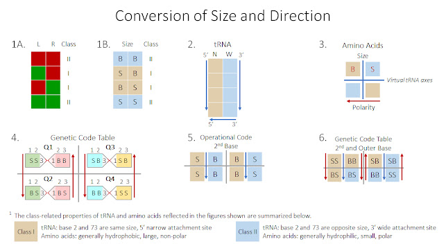
Let’s examine the implications of this restatement in greater detail. In Figure 1A, we’ve reorganized the rules table shown in Figure 2 of the previous page to make it visually apparent that two small bases (in red) or two large bases (in green) in the determinative positions (Base 2 left (L) of the vertical axis and Base 73 right (R)) are interpreted as Class II. Mixed combinations of large and small size, regardless of left-or-right position, are interpreted as Class I. The result is two combinations each of Class I and Class II components.
In Figure 1B, we’ve simply swapped the size-related red and green color codes to reflect the colors we’ve been using to designate aminoacylation class: the two same-size bases (large or small) are Class II, colored in blue. The two opposite-size bases are Class I, colored in tan. To distinguish between designations for “left” and “large,” we’ve again used “B” for “big” and retained “L” to designate “left.” In making this swap, a left-right distinction relative to the vertical axis of tRNA is reinterpreted as a distinction based on size. In both figures, four combinations of either left-right or large-small are merged into two classes.
Now we’ll look more closely at how this left-right / large-small correspondence is achieved with respect to the groove size of tRNA. Recall that when the 1D tRNA transcript folds into the 2D cloverleaf configuration, it creates a hairpin turn at the position of the embedded 3’-5’ anticodon at the bottom of the molecule. The result is two antiparallel strands that allow 5’-3’ codons on the left of the newly established vertical axis to align with their corresponding 3’-5’ anticodons on the right. On both sides of the molecule, the antiparallel strands point in the same downward direction, toward the position of the embedded anticodon at the bottom of the axis as shown in Figure 2. On the left, consecutive bases run in the 5’-3’ direction; on the right, their paired bases run in the opposite 3’-5’ direction. This common directionality on both sides of the folded tRNA structure is the logical means by which a correspondence between left-right axial placement and large-small groove size is achieved. From there, the spiraling of base pairs discussed in the previous chapter creates the alternating wide and narrow groove pattern of the double helix, each associated with one of the two sides of tRNA. In Figure 2, the left 5’ side is narrow and the right 3’ side is wide.
Having established a linkage between the large / small size of base nucleotides, the left / right sides of the tRNA axis, and the wide / narrow grooves of the tRNA molecule, let’s briefly turn to their linked component of translation – amino acids. As we described in Chapter 7, the two classes of amino acids vary with respect to size, polarity and response to the presence of water. In general, Class I amino acids are large, non-polar and hydrophobic (resistant to water) at their rear carboxyl end (right of the central alpha carbon atom in our 2D depictions). Their Class II counterparts are small, polar and hydrophilic (attracted to water) at their front amine end (left of the alpha carbon). 4
As the one-dimensional chain is being formed, all amino acids are oriented in the same direction with the amine end of one molecule attached to the carboxyl end of its neighbor immediately in front of it in the left-facing chain. However, as it undergoes secondary and tertiary folding into the final protein shape, the opposing polarity of the two classes manifests as an inside vs. outside position with respect to the final protein shape. Large Class I amino acids tend to cluster in the interior with their hydrophobic carboxyl ends closest to the center and their amine ends pointing outward. Small Class II amino acids are generally found on the protein surface, again with their carboxyl ends closest to the center and their hydrophilic amine ends pointing outward into the surrounding cellular matrix. Notice that both classes of amino acids are oriented in the same direction with their amine ends pointing outward. The distinction is their relative position within the finished protein: the amine end of large molecules clustered in the core point outward toward small molecules whose amine ends also point outward to form the surface of the protein. This unidirectional feature of the two classes of amino acids allows polarity to serve as a consistent directional reference whether it applies to an individual amino acid or to the folded protein as a whole.
During aminoacylation, Class I amino acids approach the narrow groove at the 5’ end of the folded tRNA molecule. Their Class II counterparts approach the wide groove at the opposite 3’ end. In thinking about this, it’s helpful to picture the tRNA in its cloverleaf configuration, with the biased 3’ end of the transcript clearly longer than the 5’ end.
Figure 3 is a schematic representation of the way in which these characteristics of the two classes of amino acids map to the modern structure of tRNA. In this figure, the blue lines represent the virtual axes of the tRNA molecule; the vertical line divides the 5’ side (left) from the 3’ side (right). The horizontal axis divides the top half of the molecule containing the amino acid acceptor stem from the lower half containing the embedded anticodon. The blocks represent the two classes of amino acids color-coded as before: large Class I amino acids are in tan, and their small Class II counterparts are in blue. Here, the “B” and “S” designations in red font refer to the size of the amino acids – not bases. The red arrow at the bottom of the figure serves a dual purpose. It represents the specific direction of all amino acids, with the forward amine end pointing left and the rear carboxyl end on the right. It also represents amino acid polarity and position within the folded protein, with areas of highest polarity on the left and those with lowest polarity on the right.
This overlay of amino acid size and polarity on the structural axes of the tRNA molecule shows how corresponding features of these two components of translation relate to one another. In the top half of tRNA, we see the relative size of an amino acid side chain matched to its opposite tRNA groove size; in the bottom half, we see the relative polarity of the two ends of an amino acid matched to the direction of the embedded anticodon. Let’s look more closely at how these matches are reflected in the figure. Regarding amino acid size, the top half of the figure representing the tRNA acceptor stem shows large Class I amino acids attached to the narrow groove at the 5’ end of tRNA (left in the figure), and their small Class II counterparts attached to the wide groove at the 3’ end (right). With respect to polarity, the bottom half of the figure representing the anticodon arm of tRNA shows hydrophilic Class II molecules associated with greater polarity on the left just before the turn at the position of the anticodon, and hydrophobic nonpolar Class I molecules on the right just after the turn. In the bottom half of the figure, the placement of Class I and II amino acids on either side of the vertical tRNA axis is not intended to reflect the groove attachment preference of the two classes. Here, placement on either side of the vertical axis is strictly a reflection of relative polarity as indicated by the red arrow. If we imagine the two blocks at the bottom of the figure as two individual amino acids, the amine end of a small hydrophilic amino acid would be farthest left and the carboxyl end of a large hydrophobic amino acid would be farthest right.
Considering the features of this figure more broadly, notice the correspondence between the size and direction / polarity of amino acids and the size and direction of base triplets embedded in tRNA. At the top of the figure, the size and polarity differences associated with each of the two amino acid classes are linked to an attachment preference for one of the two grooves of tRNA (wide or narrow). Large, non-polar amino acids attach at the 5’ narrow side and small, polar amino acids attach at the 3’ wide side. At the bottom of the figure, the one-directionality of individual amino acids (amine end left, carboxyl end right) corresponds to one-directional positional clustering based on polarity within the finished protein (polar Class II amino acids at the surface, non-polar Class I counterparts at the center). These orientations correspond to the one-directionality of the antiparallel strands in the folded tRNA molecule, in which both the 5’ end and the 3’ end of the folded molecule on either side of the virtual vertical axis are directed downward toward the specific 3’-5’ direction of the anticodon at the bottom of the hairpin turn.
Now that we’ve seen how size, direction and class are correlated within tRNA and amino acids, we need to explore how they are reflected in the genetic code. To do this, we’ll briefly return to the genetic code table discussed earlier in this chapter. As a reminder, this table is derived by first sorting codon triplets by the size of their second base beginning with small uracil in Column 1, and then doing a secondary sort by the size of the first base of each triplet, again beginning with uracil in Quadrant 1, Column 1. Since all triplets in the table are either codons or reverse codons, all are shown in 1-2-3 order reflecting their 5’-to-3’ direction. Organized in this way, the 32 directionally-sized couplets result in a table that can be summarized by quadrant as reflected in Figure 4 and below. This is the same table we saw in our discussion of Base Size and Codon Direction.
1-2-3 1-2-3 1-2-3 1-2-3
S-S-3 1-B-B | S-B-3 1-S-B
B-S-3 1-B-S | B-B-3 1-S-S
Now let’s see how this table maps to the directional size of tRNA as reflected in wide and narrow grooves on either side of the virtual tRNA axis and the directional size of amino acids reflected in molecular size and polar direction. Figure 5 shows the size of the second bases in each quadrant of the genetic code summarized in Figure 4; the small bases in Columns 1 and 4 are designated “S”, and the large bases in Columns 2 and 3 are designated “B”. Gray lines separate the groupings into the four quadrants as before.
Within each quadrant, the two codons are color-coded by class depending on whether they lie on the left or right side of the virtual tRNA axis as indicated by the blue arrows that lie between them. Although this means of designating class does not reflect the 3:1 / 1:3 ratio in the two halves of the genetic code table, we can legitimately use this convention as a means of organizing codon class within it because the 64 codons it contains are evenly divided with respect to class: 32 code for Class I amino acids that attach to the 5’ end on the left side of the tRNA axis (Columns 1 and 3 colored in tan), and 32 code for their Class II counterparts that attach to the 3’ end on the right side of the axis (Columns 2 and 4 colored in blue). In other words, the correlation between class designation, the left-right sides of the tRNA vertical axis and tRNA groove size outlined above allows us to convert tRNA left-right to amino acid class. 5
Here, we’ve added the size of the first base in Columns 1 and 3 beginning with small uracil in Column 1 and matched it with its opposite-sized third base in Columns 2 and 4. This gives us the same directional size relationships reflected in the genetic code table of Figure 4. Notice that while the middle base size is the same in both the upper and lower quadrants of every column in Figures 4 and 6, the outer base size is reversed. Specifically, in Columns 1 and 3, the small first base in the upper two quadrants is reversed to large in the lower quadrants; in Columns 2 and 4, the large third base in the upper quadrants is reversed to small in the lower quadrants. This reversal is indicated in both figures by the red vertical arrows nearest the base where the reversal has occurred. Since this table is a summarization of the complete genetic code table depicted in our discussion of it earlier in the chapter, it reflects the same 3:1 / 1:3 class-based asymmetry that is built into the directional size codon table. Here as in the codon table, we see this asymmetry expressed in both the top / bottom and left / right halves of the figure. 7
1 https://en.wikipedia.org/wiki/Opcode
2 https://www.openaccessgovernment.org/article/trna-the-operational-rna-code-and-protein-folding/172255/
3 Consistent with our theory of living awareness, biochemistry is a metaphor for life; the database is an expression of the truth of life.
4 These statements with respect to amino acid polarity and class are broad generalizations. Amino acid polarity does not cleanly divide by class. Some Class I amino acids, such as glutamic acid and lysine, are polar and some Class II amino acids, such as glycine and alanine, are non-polar.
5 The 32 / 32 distribution of codons by class is ideal. There are, and have been, myriad organisms with variations of this even distribution by class.
6 Since the first and second bases of a given 5’-to-3’ triplet pair with the third and second bases of its reverse codon, respectively, adding the first and third base designations in paired columns of the table establishes the 3’-5’ direction of the anticodon relative to any given 5’-3’ codon.
7 Again, it bears repeating that throughout this discussion, we are showing the relationship between codon / amino acid / tRNA classes in general; we’re not attempting to explain the relationship between individual codons and specific amino acids. With respect to the table in Figure 6, individual codon-amino acid group conversions generally fit their corresponding quadrants according to the base in the third position of the codon as shown in our discussion of the way in which amino acids are embedded in the genetic code. Refer to “Amino Acids Aren’t Outside the Code – They’re Embedded in It” and “Class Division and the Genetic Code Table” earlier in this chapter for a more complete discussion of the table summarized here.

No comments:
Post a Comment